sample recovery
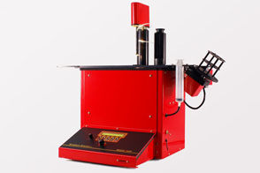
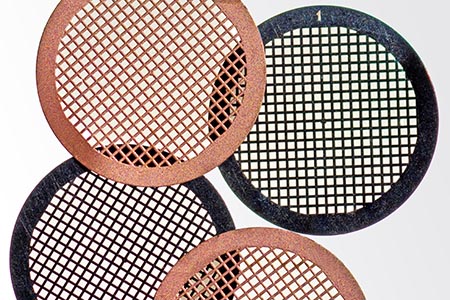
For cryo-electron microscopy (Cryo-EM), cell and tissue structures are preserved largely artefact-free, whereby all physiological processes are stopped by rapid freezing and maintained close to life. Cryo-EM requires samples no thicker than 200µm. Anything larger cannot be vitrified by high-pressure freezing. Samples investigated by cryo-EM include tissue sections, small organoids, bacteria, cells, viruses, organelles and macromolecules. Cells and tissue can be cultivated directly on TEM grids, for that the carbon films on the grids should be hydrophilized by a plasma cleaning / glow discharge. Organelles, viruses and macromolecules, on other hand, should be enriched before freezing. This works very well by using density gradient ultracentrifugation (DGUC) and gradient fractionation (BioComp Fractionator). A special method for single particle preparation is GraFix (gradient fixation). This is used to stabilise fragile macromolecule complexes by weak chemical cross-linking during DGUC (e.g. with glutaraldehyde or formaldehyde).
