Fluorescence Microscopes
(1)Fluorescence microscopy is a special method of light microscopy based on the physical effect of fluorescence. Proteins or fluorescent dyes are excited with light of a certain wavelength and emit light of a different, longer wavelength.
So-called fluorochromes (e.g. fluorescently labeled antibodies and nucleic acids or naturally fluorescent proteins) selectively bind to target structures in living or fixed samples (e.g. in cells) and make them visible under a fluorescence microscope.
The advantages of fluorescence microscopy over other techniques include:
- High resolution: fluorescence microscopes enable the visualization of cellular structures with high accuracy.
- Specific labeling: Targeted labeling of molecules or cell structures.
- Live cell imaging: Investigation of dynamic processes of living cells in real time.
- Multichannel: Using multiple dyes simultaneously.
- Quantitative analysis: Measurement of intensities.
Important areas of application for fluorescence microscopy are:
- Biology and medicine: examination of cells, proteins, gene expression and cell activity.
- Diagnostics: Immunofluorescence for the detection of antigens with antibodies in tissue samples.
- Materials science: Characterization of material samples and inclusions.
-
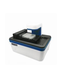 Art.Nr: NE-JS1000S
- Multi-channel fluorescence imaging
- Compact and compatible with standard CO2 incubators
- Fully automated X-Y-Z stage
- Easy and powerful software
- Capture and analyze images in real time