JuLI™ Stage - Real-time Cell Imaging and Analysis System
NE-JS1000S
- Multi-channel fluorescence imaging
- Compact and compatible with standard CO2 incubators
- Fully automated X-Y-Z stage
- Easy and powerful software
- Capture and analyze images in real time
Product Details
Description
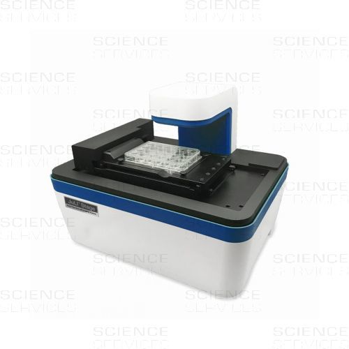
JuLI™ Stage is a digital Multifluorescence Microscope and Real-Time Cell History Recorder (CHR) designed for live cell imaging and analysis of various cell culture vessels. With its dimensions, it fits into any typical incubator.
With JuLI™ Stage, imaging, videos and editing can be done simultaneously and automatically with an intuitive set-up, software and hardware.
JuLI™ Stage is equipped with a fully automated x-y-z stage to locate and fix an area of interest within your cell culture. Multi-channel fluorescent colours can be recorded to acquire cell images and videos. Furthermore, users can obtain quantitative cell confluence results with low variation as well as growth curves using image based analysis.
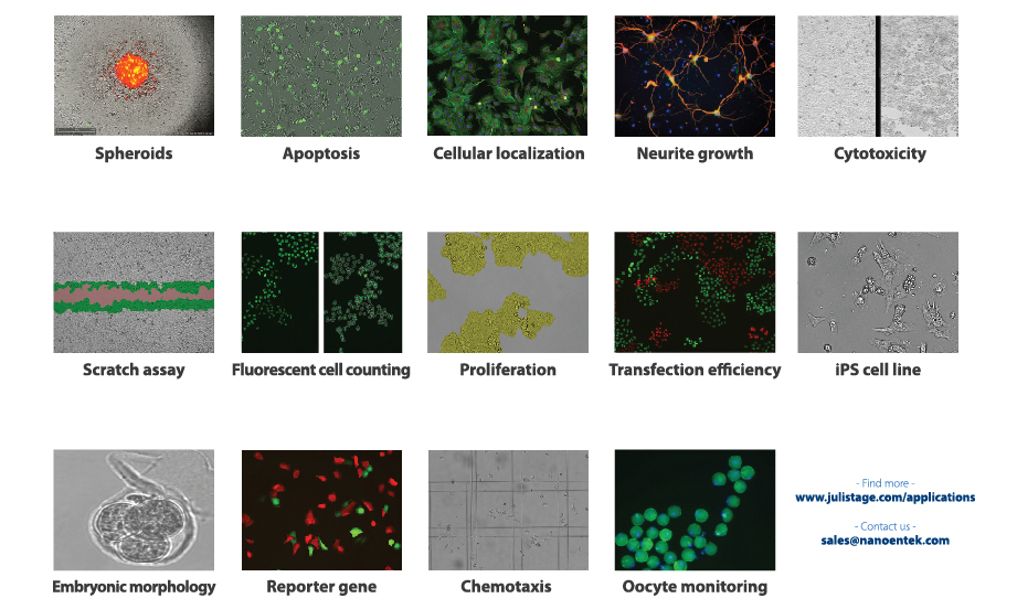
The main assays available with JuLI™ Stage are: Cell growth monitoring and analysis simultaneously in 1-384 wells and cell culture flasks (long time monitoring over 3 days), Cell proliferation, migration and differentiation, Stem cell monitoring, Wound healing (Scratch-wound assays), Apoptosis & Cytotoxicity, monitoring and analysis of fluorescence expression and subcellular localisation of recombinant proteins, 3D spheroid culture, Angiogenesis and more...
Cell lines tested so far:
- Attached cell lines: U2OS, HeLa, NIH3T3, HepG2, MCF-7, PC-12, HUVEC
- Primary cells: hMSC, Mouse oocyte stem cells, PBMC (Peripheral Blood Mononuclear cells), Neuronal stem cells
- Embryonic cell lines: Mouse embryonic cells
Main Functions:
- Cell Imaging System: real-time observation and recording of living cells and its history from beginning until the end, revert to any time point, safe time with time-lapse function
- Time-lapse recording
- Automated video making
- Image stitching: acquire a whole well by using stitching options
- Z-stack Imaging: high quality images by using the Z-stack focus option
- Image edit: Image Editor (improving image quality with background correction and auto adjust function) and Movie Maker (making various types of movies) import, verify, review and re-edit project data
- Multi-channel fluorscent colors (GFP, RFP, DAPI)
- Image Statistics Multi-well monitoring (up to 384wells)
- Multi-position monitoring: take any number of images of any positions of a well
Optional Software:
- Scratch Stat (creation of uniform scratch lines for 96-wells, real-time analysis of scratch closure)
- Spheroid Stat (various real-time analysis functions for spheroids in 96-wells)
Features
- Incubator-compatible (device will be delivered surface sterilised, please clean cables and equipment with 70% EtOH before use).
- Fully automated X-Y-Z stage
- Interchangeable objective lens (4x, 10x, 20x)
- Manual & autofocus
- Compatible with various plates & flasks (clear bottom microplates (6well, 24well, 48well, 96well and 384well), T25 and T75 culture flasks, glass/plastic Petri dishes (35mm, 60mm, 100mm)
- Data management with all-in-one PC
Applications
Specifications
| Light source | Blue, Green, UV LED (Intensity adjustable) |
| Objective lens | 4X, 10 X, 20 X + Digital ZoomSpec. |
| Fluorescence | 3 fluorescence DAPI: Excitation 390/40, Emission 452/45 GFP: Excitation 466/40, Emission 525/50 RFP: Excitation 525/50, Emission 580LP |
| Camera | High-sensitivity monochrome CCD (Sony sensor 2/3”) 1,936 ⅹ1,456 pixels (2.8 M), 53 FPS, 14 bit |
| Stage | Automated X-Y-Z stage Inter-changeable vessel holder |
| Exported formats | Image: JPEG, TIFF, BMP, PNG Video: AVI Raw data: CSV |
| PC requirement | All-in-One touch screen desktop CPU: Intel Core i5-4590S Processor (Qual Core, 6MB,3.00GHz) OS: Genuine Windows 8.1 64bit (ENG) RAM: 8 GB (2x4 GB) 1600MHz DDR3L Memory HDD: 1TB 2.5” SATA (5,400 Rpm) 23” Full HD (1920 X 1080) with touch screen |
| Operating power | 100 – 240 V, 1.5 A, 50/60 Hz |
| Electronic input | 12VDC, 5.0 A |
| Dimensions | 429 (W) X 310 (D) X 324 (H) mm |
| Weight | 18.0 kg / 39.7 lbs |
| Operating environment | 5 - 40 °C, 20 – 95 % |
| Contrast methods | Fluorescence and transmitted light |
Please find more reference papers at the NanoEnTek site >>Papers
More Information
| Electrical Power | 100 – 240 V, 1.5 A, 50/60 Hz |
|---|---|
| Electrical Supply | 12VDC, 5.0 A |
| Dimensions | 429 (W) X 310 (D) X 324 (H) mm |
| Weight | 18.000000 |
| Manufacturer |
NanoEnTek
|
