CryoCapsule®
Product Details
Description
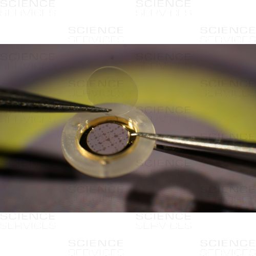
CryoCapsule®
Bringing Correlative light and electron microscopy forward with the CryoCapsule®
 High pressure freezing is the most advanced technology when it comes to vitrify a hydrated biological specimen while preserving the ultrastructure. Developed in the 80's [1], the technology evolved progressively to become accessible to a larger community. Still, the sample preparation prior to HPF remains tedious and often comes to advance expertise depending on the specimen [2].
The CryoCapsule® is a new tool in the field of High Pressure Freezing (HPF) and correlative light and electron microscopy (CLEM). Comparable to a small petri dish, it is composed of a landmarked sapphire disc and a gold spacer ring (50µm thick) maintained together by a plastic ring [3].
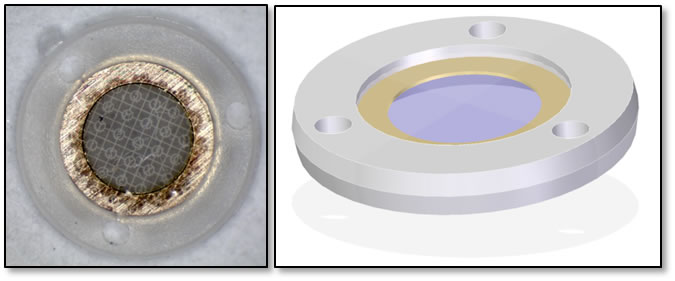
CryoCapsule® 3mm, #E3750
The specimens are encapsulated between the support sapphire disc (carbon landmarked) and a covering sapphire disc.
The CryoCapsule® is loaded into a specific adaptor (HPM010, HPM100, HPF compact 02). Live cell imaging is done directly on the specimen in the CryoCapsule® prior to HPF.


HPM010 Abra Fluid Adapter, #E3751 HMP100 Leica Adaptor, #E3752
Post-HPF, the specimen is processed for freeze substitution [4] and room temperature sectioning.
CryoCapCell has developed a set of tools to manipulate the CryoCapsule® [4] in most scientific environments.


Décapsuleur, #E3753 Décapsuleur Pencil, #E3754
References
- Moor H, Bellin G, Sandri C, Akert K. The influence of high pressure freezing on mammalian nerve tissue. Cell Tissue Res [Internet] 1980 [cited 2013 Jul 24];209:201-16.
- Mcdonald KL, Schwarz H, Thomas M, Müller-Reichert T, Webb R, Buser C, Morphew M. "Tips and tricks" for high-pressure freezing of model systems. Methods Cell Biol [Internet] 2010 [cited 2011 Apr 19];96:671-93.
- Heiligenstein X, Heiligenstein J, Delevoye C, Hurbain I, Bardin S, Paul-Gilloteaux P, Sengmanivong L, Régnier G, Salamero J, Antony C, Raposo G. The CryoCapsule®: Simplifying Correlative Light to Electron Microscopy. Traffic [Internet] 2014 [cited 2014 May 14];15:700-16.
- Heiligenstein X, Hurbain I, Delevoye C, Salamero J, Antony C, Raposo G. Step by step manipulation of the CryoCapsule® with HPM high pressure freezers. Methods Cell Biol [Internet] 2014 [cited 2014 Nov 27];124:259-74.
More Information
| Form |
round
|
|---|---|
| Material |
Saphire (synthetic)
|
| Manufacturer |
CryoCapCell
|
