3D Cell Culture, Organoids and Organ on-a-Chip
 MSCs on Hydroxylapatit Scaffolds with PI & FITC / Axiovert 5 / iHMC Embryo / Tobacco Protoplasts / Zeiss Axiovert 5
A wide range of studies over more than two decades show that 3D cell culture technology can bridge the gap between 2D in vitro cell culture and animal (in vivo) experiments. For more than 50 years, 2D cell culture has been in use in the form of monolayer or suspension cultures. Recently, there is a shift towards 3D culture technology which is proceeding steadily.
3D cultures include:
- Organoids
- Tissue engineering scaffolds (3D skin models, hydrogels)
- Organ on-a-chip (microfluidic technology)
- 3D printed tissues
Improved procedures for standardizing 3D cultures make their use in cell biology research, translational medicine, drug screening, and toxicity testing, more applicable and allows avoiding animal testing models.
The development process of a 3D culture can take different lengths of time depending on its complexity. Therefore, continuous monitoring is a prerequisite for successful establishment and documentation.
The production and investigation of 3D cultures requires both new and adapted methods in imaging as well as in assay development.
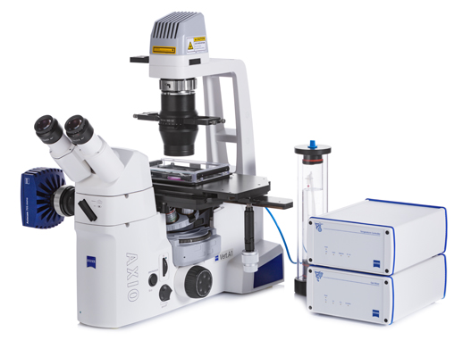
During the production process of a 3D cell culture, an inverted live cell routine microscope, such as the Axiovert 5 from ZEISS, is a reliable companion for documentation and control. It allows the imaging of three-dimensional cell cultures by using special contrast techniques, such as differential interference contrast (DIC) also for plastic vessels (PlasDIC) or Hoffmann modulation contrast (iHMC). Sample-gently LED fluorescence, the stand-alone cameras AxioCam 202/208, as well as ZEN lite and the new Labscope Multi-Channel software perfectly round off the monitoring of 3D cultures. In addition, microfluidic systems, incubation units or micromanipulators can be added to the Axiovert 5 stage.
|
Literature on 3D Cultures, Organoids and Organs on-a-Chip: |
|
Product Information |


